Tuesday, March 31, 2009

Stem cell treatment of Macular Degeneration

Below is an article from the xCell Center's website http://www.xcell-center.com/treatments/diseases-treated/macular-degeneration.aspx. In case you didn't know, Ocular Histoplasmosis Syndrome (OHS) is very similar to Wet Age-Related Macular Degeneration (AMD). AMD typically impacts people 55 and older, but AMD & OHS are treated the same b/c its the same thing happening in both cases ... just for different reasons. 

New treatments involving the retina are typically conducted on AMD patients b/c their condition can be proven. OHS is very hard, if not impossible, to prove b/c the fungus is not present ... OHS patients wait for testing to be done on AMD patients, and then we undergo the same procedures and typically get the same results. Unfortunatley, its usually not covered by our insurance as they like to see factual proof of a condition. Regardless, this break through in stem cell research is a very big step forward for both AMD and OHS patients.

The article: 

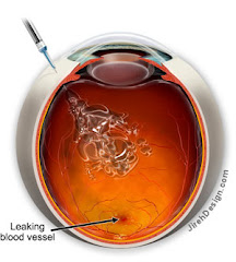
Macular degeneration is a retinal degenerative disease that causes progressive loss of central vision. The risk of developing Macular degeneration increases with age. The disease most often affects people in their sixties and seventies. Macular degeneration is the most common cause of vision loss in individuals over the age of fifty.

The macula is the central portion of the retina responsible for perceiving fine visual detail. Light sensing cells in the macula, known as photoreceptors, convert light into electrical impulses and then transfer these impulses to the brain via the optic nerve. Central vision loss from Macular degeneration occurs when photoreceptor cells in the macula degenerate.

The Xcell-Center Macular Degeneration treatment
The stem cells are first collected from a patient’s bone marrow, extracted from the hipbone (iliac crest) then implanted back into the body days later. Prior to re-implantation of the cells, the bone marrow is processed in one of our labs, where the quantity and quality of the stem cells is also checked.

These re-injected stem cells have the potential to transform into multiple types of cells and are capable of regenerating damaged tissue. Our innovative stem cell treatments use the self-healing potential of each patient’s own body to stimulate regeneration or repair.

We believe that the retro-bulbar injection of autologous stem cells might first prevent the progression of dry and wet macular degeneration by reducing the Drusen deposits in the retinal pigment epithelium beneath the macula. It might also prevent the destruction of the photoreceptors in both the dry and wet type, by reactivating proper micro-vessel activity and reducing the abnormal blood vessel growth beneath.

Results
The following results are for one female patient, 45 years of age.

In the past, she had undergone several photodynamic laser macula treatments and two "lucentis" intra vitreo injections with a disastrous clinical result. Her clinical condition continued to worsen.

When she arrived at the Xcell-center for the first time, she had a central scotoma on the left eye and she was not able to count her fingers. In the right eye we observed the onset of macula degeneration with macula epiteliopathy and 9/10 corrected visual acuity that we have been able to preserve until the present.

After some detoxification treatments the patient was sent to the XCell-center on April 25, 2008 for a bilateral autologous stem cell retro-bulbar implantation.

Just two weeks after the treatment, during an objective, standardized examination, this patient showed significant improvement.

The next picture shows the Threshold Fovea Map. Clearly visible is the improvement in retinal sensitivity.


The following picture shows the Visual Fields. Here, too, one can see an improved retinal sensitivity.


Monday, March 9, 2009

Update on my Eyes ...

I thought I'd give a quick update on my eye situation. I went in about two weeks ago as I was experiencing new blind spots in my right eye one night while driving. Considering I can't see out of my left eye, this scared me pretty badly.  So I saw my retina specialist the very next morning, and after the usual scans, he didn't see any new bleeds ... which is always good.  

I'm not too sure what I experienced the night before, but we are moving ahead cautiously as he still thinks Olivia is a little too young for me to start treatments again.  I have faith that if it comes down to that, God will take care of her ... but we are doing what we can to calm my life down so I get more rest and keep stress at bay.  

I went in last Friday again for another scan, and things still look okay. When this happened last year, I saw the spots first and then several weeks later, the scans picked them up. Its hard to rely on the scans b/c once a leak starts, it moves pretty quickly and so does my vision loss/distortion. I'm having major issues with floaters lately ... could be from the lack of sleep, but these we can gladly deal with as long as they aren't accompanied by a new leak.

So, if you'll keep my in your prayers, I would greatly appreciate it. We knew this was a risk when I found out I was pregnant. I'm so thankful that God has brought us nearly 31 weeks without any visual loss ... I'm so blessed I haven't had to choose between my sight and my daughter. 

Thank you for all your cards of support and prayers over the past couple of weeks. They really mean a lot to me and keep me encouraged!  I joke with the techs each month that Olivia will know them as her family ... I think they are as excited about her arrival as we all are.

~ Rebecca

Thursday, September 18, 2008

Been a while ...

Well, its been a while since I've posted on here. Just an update ...

I didn't need a treatment last month which was wonderful! I haven't had a leak in my right eye since January. We've just been treating for safety's sake the past several months. Dr. Gindzen says my right eye look great. God has truly answered so many prayers! (My left eye is actively leaking but since I have a permanent scar from the laser treatments, we aren't going to be treating the leak).

The VISION 2008 seminary was wonderful! Steve and I had a great time and learned so much about retinal diseases. We met some great people and their stories were so encouraging. I really has a new outlook on my condition after the conference.

Another recent development is that I'm 7 weeks pregnant. I have to tell my RS tomorrow and I know he'll be disappointed. We were supposed to wait a couple of years for a better treatment ... one which could be administered while pregnant. Well, sometimes God has different plans and this was one of them. I'm praying that God will take care of my eyes and give us a healthy baby. I know if there is one cure out there for me, its God. I have full faith that He will prevail over this disease.

Please keep us in your prayers as we go through the next 33 weeks.

~ Always in Christ, Rebecca

Wednesday, July 2, 2008

VISIONS 2008 Convention in Washington, DC

On August 8-10, Steve and I have the opportunity to go to the Vision 2008 Retinal Disease Convention in DC. What a blessing from God! Below is the link to the website if you'd like to attend or learn more about it.

http://www.blindness.org/VISIONS/


Some of the Doctors Speaking at VISIONS 2008 Include:
David Gamm, M.D., Ph.D., University of Wisconsin Hospitals and Clinics
William Kimberling, Ph.D., National Usher Syndrome Center
Janet Sunness, M.D., Greater Baltimore Medical Center
David Birch, Ph.D., Retina Foundation of the Southwest
Jacque Duncan, M.D., UCSF School of Medicine
J. Jill Hopkins, M.D., Retina-Vitreous Associates
Byron Lam, M.D., Bascom Palmer Eye Institute
Shalesh Kaushal, M.D., Ph.D., University of Florida
Dennis R. Hoff man, Ph.D., Retina Foundation of the Southwest
Linda Lemieux, Advanced Cell Technology
Estelle Condra, Author and Motivational Speaker
Rafael Caruso, M.D., National Eye Institute
Emily Chew, M.D., National Eye Institute
Morton Goldberg, M.D., National Neurovision Research Institute
Mary Elizabeth Hartnett, M.D., UNC School of Medicine
Meira Meltzer, M.A., M.S., National Eye Institute

Tuesday, June 17, 2008

Appointment - June 13, 2008

My appointment on June 13th went very well. My last treatment was on May 1st, so I have officially gone 6 weeks without a treatment. Dr. Gindzen said my right eye looked great and that the Avastin injections worked perfectly. Now, we are monitoring the eye at 4 week intervals to make sure there is no activity nor new bleeds.

The only trouble I am having is with the vitreous. The jelly-like substance has become weakened (as I explained in my previous blog) causing floaters. The floaters are causing the vision to be very distorted making it difficult to drive and read, especially at night or when my eye is tired. I have three options when it comes to the vitreous distortion.

1) Learn to live with it as I did when I lost my central vision in my left eye.
2) Continue having the Avastin shots every 4 weeks allowing the vitreous to continue to weaken so gravity will eventually pull it down, and it won't be floating the way it is now. This process of weakening could take several years and during that time, my vision would get increasingly worse. Currently the weakened vitreous has pulled away from the back of my eye but is still too firm for gravity, so it is just floating there distorting the vision instead of being pulled to the bottom of my eye by gravity.
3) Vitrectomy surgery. I have put information about this surgery at the bottom of the blog along with a video of this surgery being done on an OHS patient while removing a subretinal scar. This surgery comes with its risks ... the worse being complete blindness in that eye from an infection (1 out of 100,000). With not having my left eye to fall back on, this is an even higher risk for me.

At this point, I have chosen to live with the distortion. It took me a while to get used to losing the vision in my left eye, but I got used to it with the help of 20/20 in my right. Fortunately, the vitreous is only affecting the peripheral vision in my right eye, so I can still see 20/20. I'm not willing to risk that until the vitreous gets worse and/or impairs my macula.

The surgery is interesting though, and if you'd like to see it, watch the video at the bottom. If blood makes you queezy, I'd suggest just reading about it.

Wednesday, June 4, 2008

June Update

Well, as of today, I've officially gone six days without a treatment since March. I guess this is a great thing b/c I don't think I've had a new bleed since not getting an injection on May 29th. I've had three treatments so far at exactly 4 week intervals and my next appointment is June 13th. So, we are trying to see where the growths are at 6 weeks instead of "pre-treating" at 4.

I'm stuck between a rock and a hard place with these treatments. The Avastin is working as far as keeping the growths away, but the side effects of the injections are causing just as much trouble as the bleed would. From what I understand, the injections are weakening the vitreous causing the jelly-like fluid to break away from the back of the eye and float into my vision line. These floaters are causing my peripheral vision to be very distorted and blurry ... the only difference being these floaters float (or jiggle) around whereas a bleed stays in one spot.

Floaters are normal in an elderly person b/c as you age, the vitreous naturally weakens. But in a 26 year old, that part of your eye is very firm. The vitreous fluid is like jelly and when it pulls away from the back of the eye, it can cause retinal tears. These tears can then lead to retinal detachment. So in essense, these injections are turning my eyes into that of a 75 year old and as far as I'm concerned, doing a much harm as good.

I'm working with my PCP & RS on getting to a doctor at the Cleveland Clinic.. We have to submit several letters of recommendation to my insurance company in order to convince them to cover the treatments out-of-network. I'm also hoping that Dr. Gindzen can convince the insurance company to cover Lucentis injections since that's the FDA drug instead of Avastin, which is what I'm currently using. Hopefully the smaller molecules will do less damage and will lessen the floaters.

Due to the floaters and vision loss from my first bleed, I'm starting to have alot of trouble driving, esp at night or when my eyes are tired. I've been invited to a luncheon by the Disability Advocates this month, so hopefully I can start getting involved with that group and advocate for better public transportation in our area of town. Its a great group in Grand Rapids, and they do alot of good work for the disable.

So, that's about it. I would love some feedback from others struggling with POHS, especially if you are dealing with floaters that won't go away. Otherwise, thank you for all your prayers and please don't stop praying for my struggle with this disease as well as others who deal with this every day.

God Bless!
Rebecca

Tuesday, April 8, 2008

2nd Treatment Down

Well, last Thursday I had my second treatment of Avastin. After the first, I told myself that with every treatment, it would get easier. Man, was I wrong. This treatment hurt worse than the first and my reaction was worse.

When I had my first intravitreal injection of Avastin, I drove home afterwards; not sure how, but I did. I also went to work the next day. It hurt to work on a computer and be in the light, but I did it nonetheless. After my treatment last Thursday, I could not open my eye at all. I'm so thankful Steve took me to my appointment b/c I would have been sleeping at the doctor's office as there was no way I could have gotten home without him. The next morning, my eye was nearly swollen shut and remained that way all day. My vision was also blurred, so going to work wasn't an option.

The good news is, so far, the treatments are working, and there is a chance that I won't have to have a 3rd treatment on May 1st. I'd be so happy if I only needed an injection every 6-8 weeks instead of every 4 ... of course, to never have to have one again would be the best!

What really gets me is that down the hall, 3 elderly people were getting the same injections. I can't imagine being 80 years old and having needles stuck in my eye. They shouldn't have to go through that.

I really pray that a new treatment is found soon. Avastin is a great temporary drug, but we need something long-term. So, to all the brilliant researchers out there, please, GET TO WORK!

Another victim to POHS

My name is Rebecca. I was officially diagnosed with Ocular Histoplasmosis Syndrome in January of 2008. I grew up in Nashville, Tennessee and had never heard of the fungus Histoplasma Capsulatum until after moving to Grand Rapids, Michigan in August of 2007.

It is my hope from this blog that I can provide information to others about this terrible disease and urge people within the "Histo Belt" to check their sight on a regular basis.

INTERESTING VIDEOS

I've added some interesting videos of different retinal surgeries. I will warn you that these can be graphic, but if you're interested in knowing what surgeries have been done and how they work, take a peek!

Vitrectomy for Vitreous Opacities (Floaters)

Floaters - What are they? How do they happen?

Removal of Subretinal Neovascular Membrane due to OHS

Vitrectomy Surgery

Vitrectomy Surgery allows the Retina Surgeon to work inside of the eye and repair damage caused by a variety of diseases. The vitreous is the jelly inside the eye and is removed during the vitrectomy. YOU DO NOT NEED YOUR VITREOUS. The vitreous is important when the eye forms as a fetus grows into a baby, but it does nothing good for you after that. The vitreous is replaced with a salt solution or gas or oil and your body replaces the salt solution or gas in the vitreous cavity over a week or two.

Some vitrectomies can be performed using very small 25 gauge instruments. Most are done with 20 gauge instrument. Once the vitreous is removed, instruments like picks, forceps, lasers, and cautery can be used inside the eye to help fix problems like macular pucker, macular hole, retinal detachment, vitreous hemorrhage, diabetic retinal disease, infection, retained lens fragments and so on.

Vitrectomy surgery in our practice is usually done with local anesthesia and sedation. Since you will be sedated, YOU CANNOT EAT the morning of surgery. You will be sedated and your eye will be anesthetized. You will be monitored by an anesthesiologist or nurse anesthetist. After surgery you will have to wear a patch and shield over the eye until your appointment the following day.

If you have a retinal detachment and your doctor puts a gas or oil to hold your retina in place, you will have to hold your head in a certain position and also look in a certain position for the surgery to work. This is also true of surgery for macular holes which requires face down positioning.

After your patch is removed during your post-operative visit, you will need to take eye drops. If during the week after surgery, your eye starts to hurt more or vision starts to decline, you should call our office since that can indicate a problem with infection or elevated intraocular pressure. If you see a new shadow after surgery you need to call so we can check to make sure you do not have a retinal detachment.

Using modern microsurgical techniques, your retinal surgeon is able to repair many macular and retinal problems that were once thought unfixable. Your physician can tell you what the success rates are for your surgery including the possibility of some vision improvement and the possibility of a lot of vision improvement.

After any eye surgery there is a risk of infection, bleeding, and retinal detachment. Also, the intraocular pressure in the eye can sometimes rise necessitating extra post-operative eye drops. Make sure to call our office if you are having any problems.

What is Histoplasmosis?

Histoplasmosis is a disease caused when airborne spores of the fungus Histoplasma capsulatum are inhaled into the lungs, the primary infection site. This microscopic fungus, which is found throughout the world in river valleys and soil where bird or bat droppings accumulate, is released into the air when soil is disturbed by plowing fields, sweeping chicken coops, or digging holes.

Histoplasmosis is often so mild that it produces no apparent symptoms. Any symptoms that might occur are often similar to those from a common cold. In fact, if you had histoplasmosis symptoms, you might dismiss them as those from a cold or flu, since the body's immune system normally overcomes the infection in a few days without treatment.

However, histoplasmosis, even mild cases, can later cause a serious eye disease called ocular histoplasmosis syndrome (OHS), a leading cause of vision loss in Americans ages 20 to 40.

Where does Ocular Histoplasmosis Syndrome come from?

Scientists believe that Histoplasma capsulatum (histo) spores spread from the lungs to the eye, lodging in the choroid, a layer of blood vessels that provides blood and nutrients to the retina. The retina is the light-sensitive layer of tissue that lines the back of the eye. Scientists have not yet been able to detect any trace of the histo fungus in the eyes of patients with ocular histoplasmosis syndrome. Nevertheless, there is good reason to suspect the histo organism as the cause of OHS.

What are the symptoms of OHS?

OHS usually has no symptoms in its early stages; the initial OHS infection usually subsides without the need for treatment. This is true for other histo infections; in fact, often the only evidence that the inflammation ever occurred are tiny scars called "histo spots," which remain at the infection sites. Histo spots do not generally affect vision, but for reasons that are still not well understood, they can result in complications years--sometimes even decades--after the original eye infection. Histo spots have been associated with the growth of the abnormal blood vessels underneath the retina.

In later stages, OHS symptoms may appear if the abnormal blood vessels cause changes in vision. For example, straight lines may appear crooked or wavy, or a blind spot may appear in the field of vision. Because these symptoms indicate that OHS has already progressed enough to affect vision, anyone who has been exposed to histoplasmosis and perceives even slight changes in vision should consult an eye care professional.

How is OHS Diagnosed?

An eye care professional will usually diagnose OHS if a careful eye examination reveals two conditions: (1) The presence of histo spots, which indicate previous exposure to the histo fungus spores; and (2) Swelling of the retina, which signals the growth of new, abnormal blood vessels. To confirm the diagnosis, a dilated eye examination must be performed. This means that the pupils are enlarged temporarily with special drops, allowing the eye care professional to better examine the retina.

If fluid, blood, or abnormal blood vessels are present, an eye care professional may want to perform a diagnostic procedure called fluorescein angiography. In this procedure, a dye, injected into the patient's arm, travels to the blood vessels of the retina. The dye allows a better view of the CNV lesion, and photographs can document the location and extent to which it has spread. Particular attention is paid to how close the abnormal blood vessels are to the fovea.

Information on systemic histoplasmosis can be obtained from:

The National Institute of Allergy and Infectious Diseases (NIAID)The National Institutes of Health
Building 31, Room 7A50
31 Center Drive, MSC 2520
Bethesda, MD, 20892-2520
Telephone: 301-496-5717
Website: http://www.niaid.nih.gov